Unveiling the Power of Medical X-ray: A Comprehensive Guide
Related Articles: Unveiling the Power of Medical X-ray: A Comprehensive Guide
Introduction
With great pleasure, we will explore the intriguing topic related to Unveiling the Power of Medical X-ray: A Comprehensive Guide. Let’s weave interesting information and offer fresh perspectives to the readers.
Table of Content
Unveiling the Power of Medical X-ray: A Comprehensive Guide

Medical imaging plays a crucial role in modern healthcare, providing physicians with invaluable insights into the human body. Among the most widely utilized imaging modalities is the X-ray, a technology that has revolutionized diagnosis and treatment for over a century. This comprehensive guide delves into the intricacies of medical X-ray machines, exploring their underlying principles, diverse applications, safety considerations, and their enduring significance in healthcare.
Understanding the Fundamentals: How X-rays Work
X-ray machines generate electromagnetic radiation, a form of energy similar to visible light but with a shorter wavelength. This radiation possesses the unique ability to penetrate various materials, including soft tissues and bone, to varying degrees. When an X-ray beam interacts with the human body, different tissues absorb the radiation differently. Dense tissues, such as bone, absorb more radiation and appear white on the resulting image, while less dense tissues, such as soft tissues, absorb less radiation and appear darker.
The process of generating an X-ray image involves the following steps:
- X-ray Generation: A cathode within the X-ray tube emits electrons, which are accelerated towards a target anode made of a high-melting-point metal like tungsten. This collision of electrons with the anode generates X-rays.
- Beam Collimation: The X-ray beam is then directed towards the patient using a collimator, a device that shapes the beam into a defined area, minimizing unnecessary radiation exposure.
- Image Capture: The X-ray beam passes through the patient’s body, interacting with different tissues as described above. The resulting radiation pattern is captured by a detector, typically a digital sensor or X-ray film.
- Image Processing: The detector converts the radiation pattern into a digital image, which is then processed and displayed on a monitor for interpretation by a radiologist.
Applications of Medical X-ray: A Wide Range of Uses
X-ray technology has become an indispensable tool in numerous medical specialties, offering a wide range of diagnostic and therapeutic applications. Some prominent examples include:
1. Diagnostic Imaging:
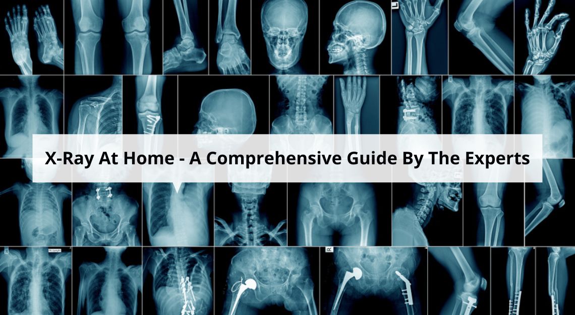
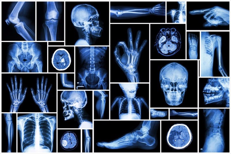
- Bone Fractures: X-rays are the primary imaging modality for detecting bone fractures, dislocations, and other skeletal injuries.
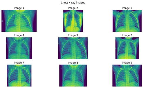
- Lung Conditions: Chest X-rays are crucial for diagnosing and monitoring lung infections, pneumonia, tuberculosis, and other respiratory illnesses.
- Dental Examinations: X-rays are used to assess the condition of teeth, identify cavities, and plan dental procedures.
- Foreign Body Detection: X-rays can help locate foreign objects lodged within the body, such as swallowed items or fragments of metal.
- Gastrointestinal Imaging: Barium studies, which involve swallowing or injecting a contrast medium, allow X-rays to visualize the digestive tract, identifying abnormalities like ulcers, tumors, and blockages.
- Cardiovascular Imaging: Fluoroscopy, a continuous X-ray imaging technique, allows visualization of the heart and blood vessels, assisting in procedures like angioplasty and stent placement.
2. Therapeutic Applications:
- Radiation Therapy: X-rays are used in radiation therapy to target and destroy cancerous cells while minimizing damage to healthy tissues.
- Stereotactic Radiosurgery: This highly precise form of radiation therapy uses X-ray beams to treat tumors and other lesions with minimal side effects.
Safety Considerations: Minimizing Radiation Exposure
While X-rays are a valuable diagnostic tool, it is crucial to recognize the potential risks associated with radiation exposure. Excessive radiation can damage cells and tissues, increasing the risk of cancer in the long term. Therefore, minimizing radiation exposure is paramount.
Minimizing Radiation Exposure:
- ALARA Principle: The "As Low As Reasonably Achievable" (ALARA) principle guides the use of X-rays, emphasizing the need to reduce radiation exposure to the lowest possible level without compromising the diagnostic quality of the image.
- Collimation: Precise collimation ensures that only the targeted area is exposed to radiation, limiting unnecessary exposure to surrounding tissues.
- Shielding: Lead aprons and other shielding materials are used to protect sensitive areas of the body, such as the reproductive organs, from direct radiation exposure.
- Image Optimization: Advanced imaging techniques, such as digital radiography, help minimize radiation exposure while maintaining image quality.
- Pregnancy Considerations: Pregnant women should avoid unnecessary X-ray examinations, especially during the first trimester, as radiation exposure can potentially harm the developing fetus.
FAQs: Addressing Common Questions
1. What are the risks associated with X-rays?
While X-rays are generally safe when used appropriately, excessive exposure can increase the risk of developing cancer. However, the risks associated with a single X-ray examination are typically very low.
2. How often can I have an X-ray?
The frequency of X-ray examinations depends on individual circumstances and the specific medical condition being investigated. Your healthcare provider will determine the optimal frequency based on your needs.
3. Are X-rays painful?
X-ray examinations are generally painless. You may experience a brief sensation of pressure from the X-ray machine, but this is usually minimal.
4. What are the alternatives to X-rays?
Depending on the medical condition, alternative imaging modalities may be available, such as ultrasound, MRI, or CT scans. Your healthcare provider will recommend the most appropriate imaging technique for your specific needs.
5. What should I do if I am concerned about radiation exposure?
Discuss your concerns with your healthcare provider. They can explain the risks and benefits of X-ray examinations and address any specific questions you may have.
Tips for Patients: Enhancing the X-ray Experience
- Communicate with your healthcare provider: Inform your doctor about any relevant medical history, including allergies, pregnancy, or any other medical conditions.
- Follow instructions: Carefully follow your healthcare provider’s instructions regarding preparation for the X-ray examination, such as removing jewelry or metal objects.
- Remain still: During the X-ray procedure, it is essential to remain still to ensure clear and accurate images.
- Ask questions: Do not hesitate to ask your healthcare provider any questions you may have about the X-ray procedure, its risks, and its benefits.
Conclusion: The Enduring Legacy of Medical X-ray
Medical X-ray technology has come a long way since its inception, evolving from a rudimentary diagnostic tool to a sophisticated and versatile imaging modality. Its ability to provide invaluable insights into the human body has revolutionized healthcare, enabling early diagnosis, accurate treatment planning, and effective monitoring of various medical conditions. While safety considerations remain paramount, the benefits of X-ray technology continue to shape the future of medicine, ensuring its enduring legacy in healthcare for generations to come.


![]()


Closure
Thus, we hope this article has provided valuable insights into Unveiling the Power of Medical X-ray: A Comprehensive Guide. We appreciate your attention to our article. See you in our next article!
